Multimodal imaging
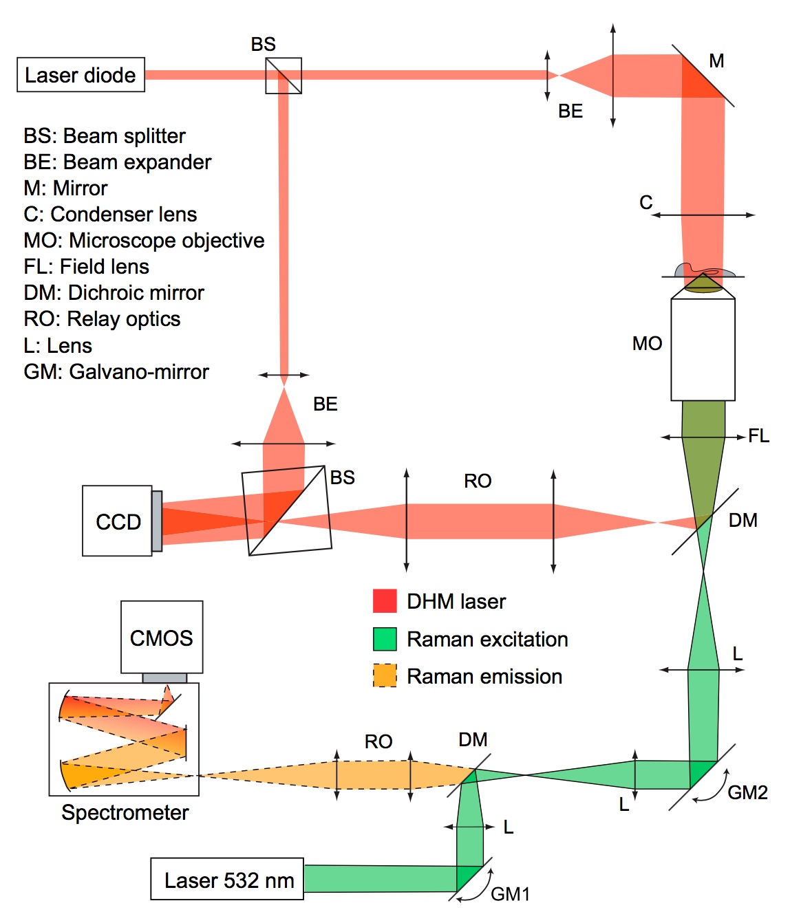
Multimodal setup that enables simultaneous Raman and quantitative phase imaging.
We developed a multimodal platform that combines several label-free imaging modalities, which enable the observation of live cell cultures without requiring any preparation protocols.
The combined label-free approaches each have individual advantages that improve the performances of the multimodal system:
- Raman spectroscopy provides insight about the local molecular content of cells, giving specificity to certain molecular species.
- Quantitative phase microscopy (QPM) enables highly non-invasive observation of cells at high speed, giving information about cellular dynamics.
The combination of these two modalities provide simultaneous information about cellular morphology, dynamics and molecular content that can be used to derive biologically relevant indicators for cellular state analysis.
The two modalities can be acquired independently and simultaneously by taking advantage of the narrow bandwidth used by QPM as it relies on interferometric measurements. The laser line of QPM is set in a range situated outside the large bandwidth of the Raman signal. This makes it possible to separate the two signals through spectral separation, as classically done for instance in fluorescence microscopy.
The two modes both rely on endogenous contrast that provides complementary information, as they rely on different physical processes. QPM measures the integrated refractive index, which derives from the elastic scattering of photons that originates from the permittivity tensor. On the other hand, the Raman process relies on inelastic scattering which is defined by the derivative of the polarizability tensor.
Mouse embryonic fibroblasts observed through simultaneous acquisition of video-rate QPM (left) during Raman acquisition (right) where the image gradually appears during laser scanning. Red: lipids (2865 cm-1), Green: C-H stretching (2973 cm-1).