
Raman imaging
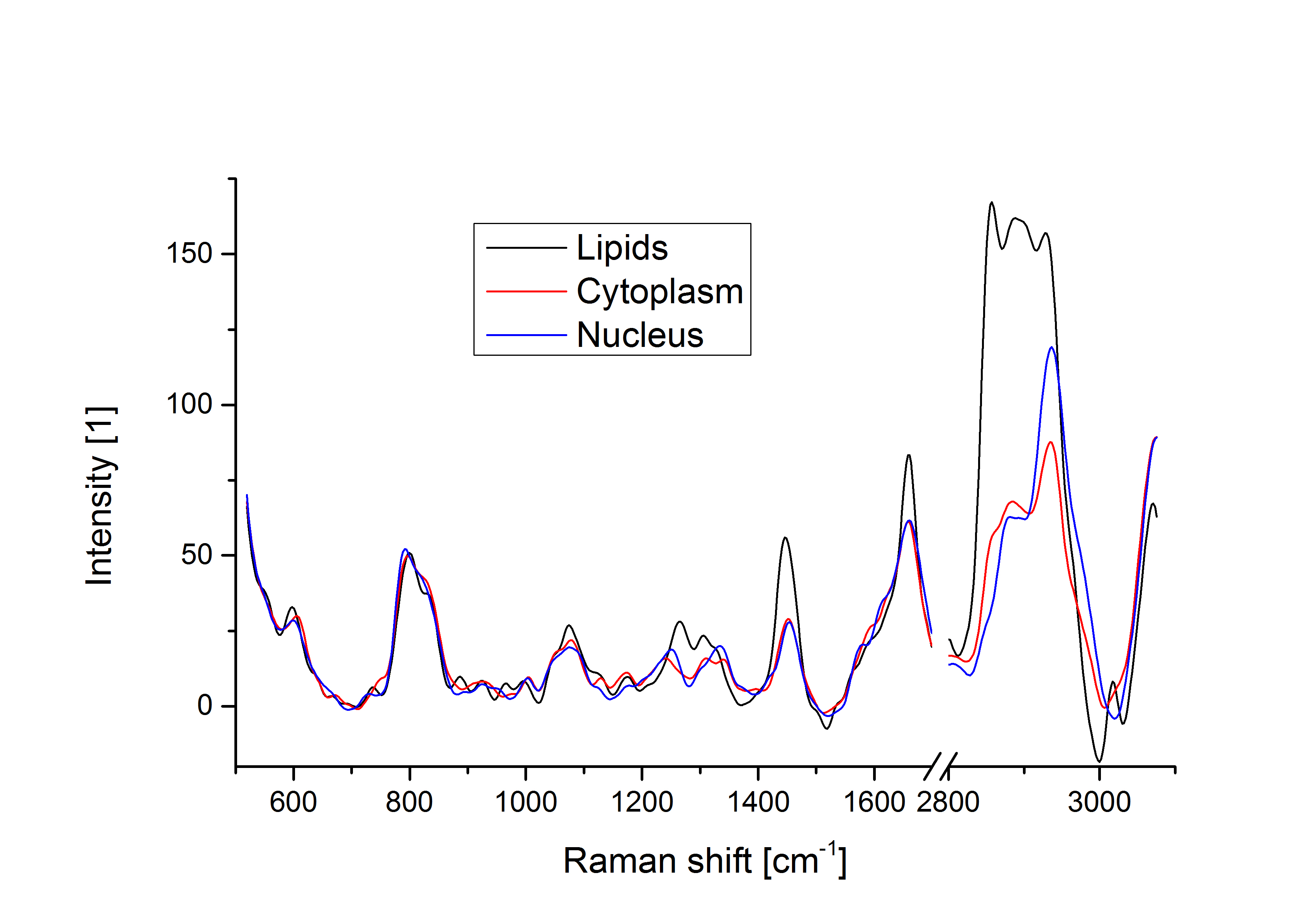
Typical Raman spectra from different regions of the cell, showing differences in molecular content.
Raman spectroscopy is a method enabling the retrieval of molecular information from a biological sample. The measurement relies on the inelastic scattering, which shifts the energy of photons based on the vibrational modes in the molecule. This produces specific peaks of red-shifted light that can be measured to identify molecules. In the case of a complex system such as a live cell, the resulting Raman spectrum is the combination of all present molecules giving a broader spectrum.
With the increase of sensitivity of digital detectors in recent years, it became possible to reduce the exposure time sufficiently to be able to perform Raman imaging by scanning the laser spot through the field of view and retrieve a spectrum for each pixel in the image, giving a hyperspectral image. The spectra at different pixels then provide molecular contrast that can highlight various features in the cell, such as lipids, nuclei, or proteins.
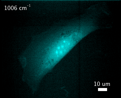
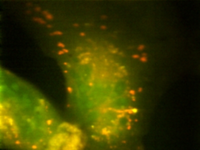
(left) Molecular images at different wave numbers obtained from the raw data, highlighting different parts of the cell. (right) Colored image obtained from the Raman data, highlighting lipids (red, 2865 cm-1) and C-H stretching (green, 2973 cm-1).